
Translate this page into:
Age estimation using cephalometric analysis of hyoid bone: A radiological study
 , Prema Anbarasu2,, Saravana Kumar Subramanian3, K. V. Kaviya1, Indra Annamalai2, K. Thrivikhraman4

*Corresponding author: Prema Anbarasu, Department of Orthodontics, Chettinad Dental College and Research Institute, Chennai, Tamil Nadu, India. prema.arasu@gmail.com
-
Received: ,
Accepted: ,
How to cite this article: Harish Raghav A, Anbarasu P, Subramanian S, Kaviya KV, Annamalai I, Thrivikhraman K. Age estimation using cephalometric analysis of hyoid bone: A radiological study. J Academy Dent Educ. 2024;10:5-9. doi: 10.25259/JADE_52_2023
Abstract
Objectives:
The study was done to estimate age using cephalometric analysis of hyoid bone. Age estimation is not only limited to forensic analysis but also to clinical dentistry. Orthodontia, one of the dental specialties, requires age estimation for the choice of treatment planning. Since cephalograms are commonly preferred for estimating age, this simple and novel method would have the added advantage of minimizing radiation exposure for the patient for the requirement of additional radiographs in the estimation of age as in hand wrist radiographs.
Material and Methods:
A total of 234 radiographs with lateral cephalometric projection were employed in the conducted study. The cephalograms were divided based on gender. Cephalometric tracing was done using hyoid bone triangle analysis established by Bibby and Preston in 1981. The triangle is created by the following: Gnathion (Gn), Retro-gnathion (R-Gn), Hyoidale (H), and C3 vertebrae were traced. The Frankfort horizontal plane served as a standardized reference in the study.
Results:
Out of all the dimensions comprising the hyoid bone triangle (C3-H, H-R-Gn, and C3-R-Gn), C3-H values were statistically significant for age estimation at P < 0.001 for both sexes. The study determined that the mean values C3-H and H-R-Gn are 35.66 mm ± 0.44 mm and 40.33 mm ± 0.54 mm, respectively. The hyoid bone is situated in a downward and posterior position.
Conclusion:
Since the certainty of the evidence was high for estimation of age, which is the need of the hour in forensics as well as in dentistry, and there is more demand for cost-effective alternatives, this method will help us reap the benefits.
Keywords
Cephalometrics
Hyoid bone triangle
C3-H
Age-estimation
Forensics

INTRODUCTION
Identification of a person relies on three general factors: Sex, age, and stature. The estimation of age holds significant importance for both living and deceased individuals.[1,2] Age estimation finds applications in forensic human identification, clinical practice, research, and legal proceedings. Various methods are employed for age estimation, including visual, morphological, radiological, and biochemical approaches. The radiological method is commonly preferred due to its nondestructive nature and simplicity.[3]
In the field of Dentistry, calculating the chronological age is necessary for orthodontic treatment planning. Since there can be individual variations in growth duration and velocity, age criteria play a key role in determining treatment options, the appropriate timing to initiate treatment, establishing prognosis, and devising a retention strategy.[4]
During an individual’s growth, bones undergo a series of changes that follow a chronological pattern, facilitating age estimation. Among these bones, the hyoid, which is a solitary floating bone in the head and neck region holds significant importance in both age estimation and orthodontic treatment planning.[5-7] The structure and positioning of the hyoid bone in the head and neck region can influence the approaches and methods employed in orthodontic treatment.[8-11]
The hyoid is linked to various intraoral and extraoral structures, including the tongue and mandible, through muscles. They play a crucial role in orthodontic treatment planning.[11,12] Furthermore, the hyoid bone is also considered important in assessing oral frailty, which is an early indicator of orofacial muscle weakness. Oral frailty can influence dental treatment and swallowing. As a result of the anatomical and physiological connections, any pathology or dimensional change in the connected structures directly influences the positioning of the hyoid bone.[12,13]
Measuring the precise position of the hyoid bone can be challenging due to its high variability, even within the same individual and position. To address this issue, the hyoid bone triangle analysis was developed. The results obtained from the hyoid triangle analysis in cephalometric radiographs, as established by Bibby and Preston, are considered more accurate compared to using other reference planes.[14]
The previous studies have focused on establishing the relationship between the hyoid bone and various growth patterns in individuals with different types of malocclusions.[13-18] Since cephalometric radiographs are commonly used for age estimation[13,19] and hyoid bone triangle analysis provides an accurate reference plane, the primary aim of this study is to estimate the chronological age of patients by employing hyoid bone triangle analysis using cephalometric radiographs. Based on our understanding, the present study presents a novel approach to estimate age that offers the advantage of minimizing radiation exposure by eliminating the requirement for additional radiographs like hand-wrist radiographs.
MATERIAL AND METHODS
This retrospective study obtained ethical approval from the Institutional Ethical Committee (Ref no: IHEC-I/0733/22). Lateral cephalograms from the Department of Orthodontics and Dentofacial Orthopedics archives spanning 10 years (2011–2020) were used for the study. All cephalograms were taken by a qualified radiographer under standardized conditions, ensuring that the F-H reference plane and the floor were parallel to each other. The same cephalogram was used for all radiographs, and the radiographic technique and exposure parameters were standardized.
The cephalograms were divided by gender, and inclusion criteria included proper positioning of subjects in the X-rays, absence of gross abnormalities, clarity of the X-ray, and retrieval of lateral cephalograms from the study period. The sample size was determined to be 234, calculated with a power (1-β) of 99%, a level of significance of 5%, and a 1% margin of error.
The lateral cephalograms were traced by the same operator using an X-ray illuminator. An independent examiner cross-checked all anatomical landmarks and tracings. Selected anatomical landmarks were used to trace planes, lines, and angles, including gnathion (Gn), the most anterior and lowermost point on the bony outline of the mandible; retrognathism (RGn), the most posterior and lowermost point on the bony outline of the mandible; hyoidale (H), the anterior superior most point on the body of the hyoid; C3 (lowermost and anterior point on the third cervical vertebrae), and the (Frankfort horizontal plane (FH plane) connecting Porion and Orbitale) [Figure 1].

- Ref points traced: C3-H, H-RGn, and C-RGn forming a hyoid bone triangle. R-Gn: Retro-gnathion, H: Hyoidale.
The hyoid triangle was traced using the above landmarks, and the measurements of the hyoid bone triangle (C3-H, H-R-Gn, and C3-R-Gn) were analyzed and included in the statistical analysis.
The measured data were recorded and analyzed using the Statistical Package for the Social Sciences Version 26 software (IBM, Chicago). One-way analysis of variance and linear regression analysis were performed, with statistical significance at P < 0.05.
RESULTS
From the measurement, the mean value of the following parameters, C3-H and H-RGn, was 35.76 mm ± 0.44 mm and 40.33 mm ± 0.54 mm. Among all the dimensions of the hyoid bone triangle (C3-H, H-R-Gn, and C-R-Gn), the distance between cervical vertebrae (C3) to hyoid bone (H) was found to be statistically significant for both age estimation and gender prediction, and the mean value of C3-H for females was recorded to be 3.57 ± 0.43 and for males 3.58 ± 0.43 [Figure 2]. Tables 1 and 2 depict the sample size, mean value, and standard deviation (SD) of C3-H and H-R-Gn. This study illustrates the mean and SD of 35.76 mm ± 0.44 mm and 40.33 mm ± 0.54 mm. The mean value of C3-H for females was recorded to be 3.57 ± 0.43, and for males, 3.58 ± 0.43. The present research confirmed a correlation between the Gonial angle of males and females, which were statistically significant. Furthermore, there is a satisfactory correlation between real age and estimated age (r2 = 0.146) [Figure 3, Tables 3 and 4].
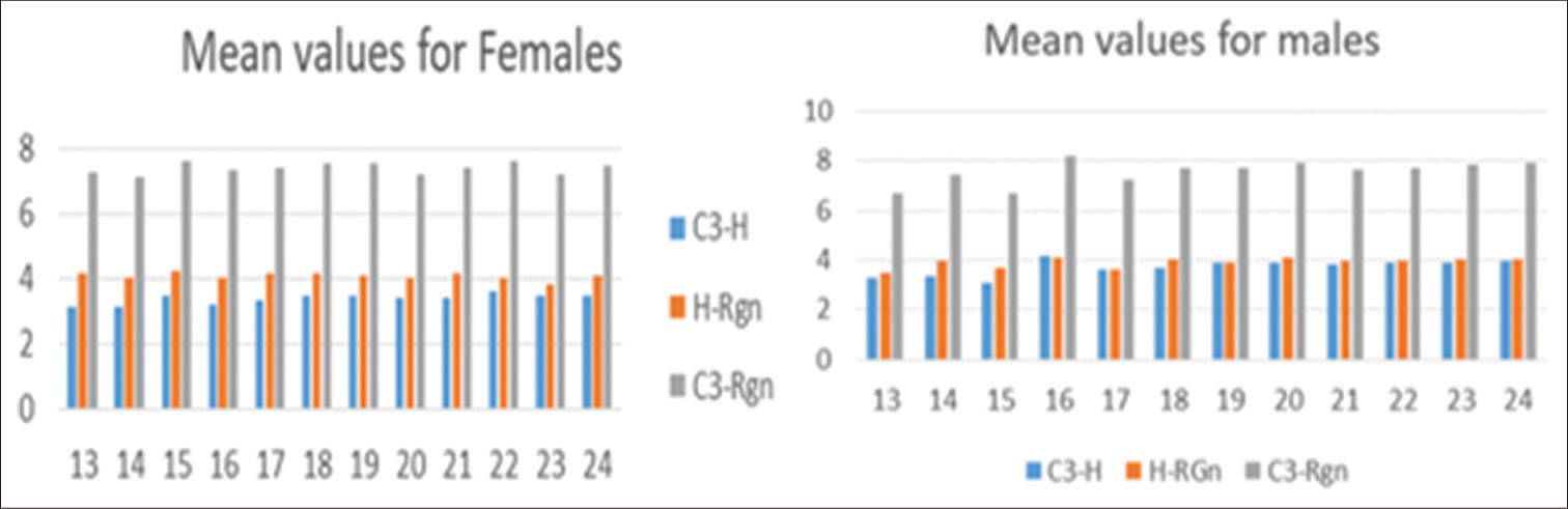
- Graphical representation of mean values of hyoid bone triangle (C3-H, H-R-Gn, and C-RGn), dimensions across age groups 13–24 for males and females. R-Gn: Retro-gnathion, H: Hyoidale.
| Mean | Std. deviation | n | |
|---|---|---|---|
| Age | 18.76 | 3.430 | 234 |
| C3-H length (in cm) | 3.576 | 0.4408 | 234 |
| H-RGn length (in cm) | 4.033 | 0.5414 | 234 |
R-Gn: Retro-gnathion, H: Hyoidale
| Age | C3-H length | H-RGn length | |
|---|---|---|---|
| Pearson correlation | |||
| Age | 1.000 | 0.382 | 0.032 |
| C3-H length | 0.382 | 1.000 | 0.232 |
| H-RGn length | 0.032 | 0.232 | 1.000 |
| Sig. (1-tailed) | |||
| Age | . | <0.001 | 0.315 |
| C3-H length | 0.000 | . | 0.000 |
| H-RGn | 0.315 | 0.000 | 0. |
R-Gn: Retro-gnathion, H: Hyoidale. sig: Significance
| Variables | Coefficients | Standard error | t-stat | P-value | Lower 95% | Upper 95% | Lower 95.0% | Upper 95.0% |
|---|---|---|---|---|---|---|---|---|
| Intercept | 2.654259 | 0.148683 | 17.85178 | 1.94 E-45 | 2.361317 | 2.9472 | 2.361317 | 2.9472 |
| Age* | 0.049135 | 0.007797 | 6.302135 | 1.46 E-09 | 0.033774 | 0.064496 | 0.033774 | 0.064496 |
| Variables | Coefficients | Standard error | t-stat | P-value | Lower 95% | Upper 95% | Lower 95.0% | Upper 95.0% |
|---|---|---|---|---|---|---|---|---|
| Intercept | −0.42149 | 0.236886 | −1.7793 | 0.0765 | −0.88821 | 0.045232 | −0.88821 | 0.045232 |
| Gender | 0.520589 | 0.065747 | 7.918116 | 9.94 E-14 | 0.391052 | 0.650125 | 0.391052 | 0.650125 |
H: Hyoidale
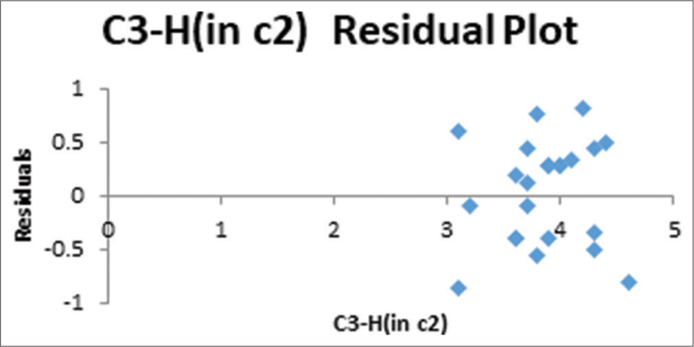
- Residual plot across C3-H and gender. H: Hyoidale.
DISCUSSION
This study aimed to determine chronological age through hyoid bone triangle analysis using cephalometric landmarks. The specific objectives were to record the basic dimensions of the hyoid bone triangle for different age groups and to compare these measurements between genders. The hyoid bone holds significance in both dentistry and forensics. Forensic experts face challenges in determining unknown facts but overcome them with innovative methodologies. While modern technologies exist, their applicability may be limited in certain situations, making non-destructive radiological methods with simpler techniques preferable. Age estimation using lateral cephalograms is commonly employed in dentistry. This study conducted a cephalometric analysis of the hyoid using hyoid bone triangle analysis to estimate age group and attempt gender identity based on it.
Despite the fact that the location of the hyoid bone is under the guidance of even small movements of the head and surrounding structures, standardization was maintained throughout the study. The H-R-Gn specification of the hyoid bone triangle is affected by the variation in the mandibular dimensions. The C3-H component showed statistical significance (P < 0.001) for both age and gender estimation. While H-R-Gn had statistically significant results for gender determination (P < 0.05), the regression analysis yielded a weak R-value (R = 0.019).
Regarding age estimation, H-R-Gn values were not significant, but the mean values of H-RGn in males remained constant above 18 years, while the same trend was observed for C3-H values in females. This could be attributed to the posterior and superior positioning of the hyoid bone in females compared to males, showing variations among different skeletal classes. An interesting finding was that there is a downward displacement of the hyoid bone with age.[20]
The overall mean parameters of the hyoid triangle were higher compared to the previous studies.[20,21] The overall mean value of the parameters selected is greater than the Pakistani populations on compared with their values. This shows that in our study population, the hyoid bone is positioned posterior and downward. The present study was conducted in the Indian state of Tamil Nadu. Further studies with larger sample sizes and expanded parameters should be conducted to investigate the effects of age, sex, and skeletal patterns on the position of the hyoid bone. The predictability of two-dimensional cephalograms for assessment of age and gender may not be very precise. With the 3D images, more predictable results can be obtained.
CONCLUSION
The study determined that the mean values of parameters used to assess the position of the hyoid bone within the study population are 35.76 mm ± 0.44 mm (C3-H) and 40.33 mm ± 0.54 mm (H-RGn). The hyoid bone is positioned downward and posterior in the studied age group. This criterion has the potential to hold significance in age determination for Medical jurisprudence, thus aiding legal investigations.
The findings from this study have implications for orthodontic treatment planning, particularly in cases involving dental and skeletal malocclusions.
Gaining a deeper understanding of the variation of the hyoid bone location among different gender and age groups can assist in diagnosing and predicting illnesses such as obstructive sleep apnea, which predominantly affects middle-aged males.
Ethical approval
The authors declare that they have taken the Institutional Ethical Committee approval and the approval number is IHEC-I/0733/22.
Declaration of patient consent
Patient’s consent not required as there are no patients in this study.
Conflicts of interest
There are no conflicts of interest.
Use of artificial intelligence (AI)-assisted technology for manuscript preparation
The authors confirm that there was no use of artificial intelligence (AI)-assisted technology for assisting in the writing or editing of the manuscript and no images were manipulated using AI.
Financial support and sponsorship
Nil.
References
- Age estimation methods using anthropological parameters on human teeth. Forensic Sci Int. 2006;162:13-6.
- [CrossRef] [PubMed] [Google Scholar]
- Dating and age determination of biological materials London: Croom Helm; 1986. p. :1-292.
- [Google Scholar]
- Nondestructive dental age calculation methods in adults: Intra and inter-observer effects. Foren Sci Int. 2002;126:221-6.
- [CrossRef] [PubMed] [Google Scholar]
- Postural change of the hyoid bone following osteotomy of the mandible. Oral Surg Oral Med Oral Pathol. 1967;23:688-92.
- [CrossRef] [PubMed] [Google Scholar]
- Forensic application of the skeletonized hyoid bone and ossified structures of the larynx in an adult American sample Tennessee: University of Tennessee; 1992.
- [Google Scholar]
- Fracture of the hyoid bone in strangulation: Comparison of fractured and unfractured hyoids from victims of strangulation. J Forensic Sci. 1996;41:110-3.
- [CrossRef] [Google Scholar]
- Age and ossification of the hyoid bone: Forensic implications. J Forensic Sci. 1987;32:1655-9.
- [CrossRef] [PubMed] [Google Scholar]
- Study of the age of fusion of hyoid bone. Leg Med (Tokyo). 2008;10:253-6.
- [CrossRef] [PubMed] [Google Scholar]
- Changes in the airway and hyoid position in response to the mandibular protrusion in subjects with obstructive sleep apnoea (OSA) Eur J Orthod. 1999;21:363-76.
- [CrossRef] [PubMed] [Google Scholar]
- Cephalometric assessment of the hyoid bone position in oral breathing children. Rev Bras Otorrinolaringol. 2007;73:45-50.
- [CrossRef] [PubMed] [Google Scholar]
- Cephalometric appraisal of the hyoid triangle in Brazilian people of Piracicaba's region. Braz J Oral Sci. 2006;5:1001-6.
- [Google Scholar]
- Hyoid bone position related to gender and aging using lateral cephalometric radiographs. J Orthod Waves. 2018;77:226-31.
- [CrossRef] [Google Scholar]
- An evaluation of upper and lower pharyngeal airway width, tongue posture, and hyoid bone position in subjects with different growth patterns. J Clin Diagn Res. 2016;10:ZC79-83.
- [CrossRef] [PubMed] [Google Scholar]
- Hyoid bone position in orthodontic patients. Orthod J Nepal. 2019;9:20-2.
- [CrossRef] [Google Scholar]
- Comparison of the changes in hyoid bone position in subjects with normodivergent and hyper-divergent growth patterns: A cephalometric study. APOS Trends Orthod. 2017;7:224-9.
- [CrossRef] [Google Scholar]
- A radiographic study of hyoid bone position in angle's class I, II, and Ill malocclusions, Master's Thesis, University of Kansas City. 1959.
- [Google Scholar]
- The position of hyoid bone in different facial patterns: A lateral cephalometric study. Eur Sci J. 2014;10:1857-81.
- [Google Scholar]
- Mandibular growth changes and maturation of cervical vertebrae: A longitudinal cephalometric study. Angle Orthod. 1988;58:179-84.
- [Google Scholar]
- Skeletal maturation evaluation using cervical vertebrae. Am J Orthod Dentofac Orthop. 1995;107:58-66.
- [CrossRef] [PubMed] [Google Scholar]
- Hyoid bone position in different facial skeletal patterns. J Clin Exp Dent. 2018;10:e346-51.
- [CrossRef] [PubMed] [Google Scholar]


