Translate this page into:
Terahertz Ray Imaging: a new ray of hope in Imaging Science
Address for Correspondence: Devika R. Krishnan Intern, Amrita School of Dentistry, AIMS Health Care Campus, Ponekkara, AIMS PO, Edappally, Cochin, Kerala-682041 E-mail: devika.diva@gmail.com
-
Received: ,
Accepted: ,
This article was originally published by Informatics Publishing and was migrated to Scientific Scholar after the change of Publisher.
How to cite this article: Krishnan DR. Terahertz ray imaging: a new ray of hope in imaging science. J Dent Educ. 2014; 1(1):51–56.
Abstract
Imaging technology is becoming increasingly prevalent in our society.Even though there were many successful imaging modalities ever since the advent of roentgen rays, they all have their shortcomings. And imaging of dental tissue had always been a challenge in imaging science due to the various layers which differ in physical and chemical and structural properties. Considerable improvisations of existing techniques and new research activities are going on in the development of newer imaging modalities that could revolutionize the science of dental radiology.
Keywords
Imaging
Radiology
Revolutionize
1. Introduction
Imaging technology is becoming increasingly prevalent in our society. Even though there were many successful imaging modalities ever since the advent of roentgen rays, they all have their shortcomings. And imaging of dental tissue had always been a challenge in imaging science due to the various layers which differ in physical and chemical and structural properties. Considerable improvisations of existing techniques and new research activities are going on in the development of newer imaging modalities that could revolutionize the science of dental radiology. Clinicians felt that fundamentally different physical principles are needed to provide safer and more cost effective imaging techniques. Thus physicists are turning into other regions of electromagnetic spectrum to address these issues.
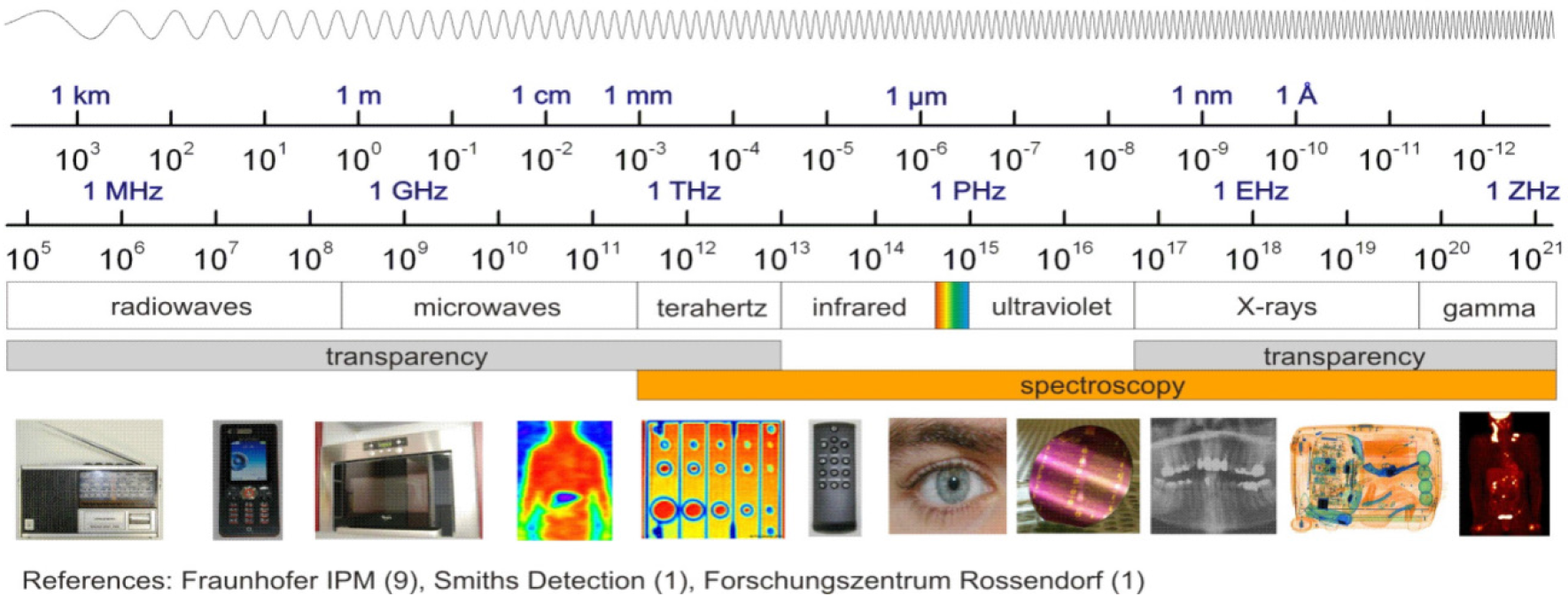
Terahertz (THz, 1THz=1012 Hz) radiation, also termed as THz waves, THz light, or T-rays typically defined as 0.1–10 THz, is sandwiched between the microwaves and the infra-red, bridging the gap between electronics and optics (Sharma, Arya & Jhildiyal, 2011). Experiments regarding T-rays date back to measurements of black body radiation using a bolometer in the 1890s. Terahertz imaging was originally developed as a research tool and is now a viable imaging and diagnostic technique.
2. Rationale
The energy level of 1 THz is only about 4.14 meV (which is much less than the energy of X-rays 0.12 to 120 keV); it therefore does not pose an ionization hazard as in X-ray radiation. Research into safe levels of exposure has also been carried out through studies on keratinocytes and blood leukocytes, neither of which has revealed any detectable alterations. This non-ionizing nature is a crucial property that lends THz techniques to medical applications (Yu et al., 2012).
Molecular interactions in the THz regime are with the protein structure. In recent years, dynamic signatures of the THz frequency vibrations in RNA and DNA strands have been characterized. In a protein-water network, the protein’s structure and dynamics are affected by the surrounding water which is called biological water, or hydration water. Hydrogen bonds, which are weak attractive forces, form between the hydrated water molecules and the side chains of protein. These affect the dynamic relaxation properties of protein and enable distinction between the hydration water layer and bulk water. The effects of the hydrogen bonds associated with the intermolecular information are able to be detected using THz spectroscopy. THz spectra contain information about intermolecular modes as well as intra-molecular bonds and thus usually carry more structural information than vibrations in the mid-infrared spectral region which tend to be dominated by intra-molecular vibrations. (Sun et al., 2011; Yu et al., 2012).
In addition to providing valuable spectral information, 2D images can be obtained with THz time-domain spectroscopy (THz-TDS) method by spatial scanning of either the THz beam or the object itself. In this way, geometrical images of the sample can be produced to reveal its inner structures. Thus, it is possible to provide three-dimensional views into a layered structure. When a THz pulse is incident on such a target, a train of pulses will be reflected back from the various interfaces. For each individual pulse in the detected signal, the amplitude and timing are different and can be measured precisely. The principle of time of flight technique is to estimate the depth information of the internal dielectric profiles of the target through the time that is required to travel over a certain distance. This permits looking into the inside of optically opaque material and it has been used for THz 3D imaging. (Sun et al., 2011; Yu et al, 2012).
3. Pros and Cons of Imaging Techniques THz Systems
The technology for generating and detecting THz radiation has advanced considerably over the past two decades. According to the laser source used, THz systems can be divided into two general classes: Continuous Wave (CW) and pulsed. It is also possible to generate THz radiation using oscillatory methods and this is often less expensive than using a laser based approach.
A typical CW system can produce a single fixed frequency or several discrete frequency outputs. Some of them can be tunable. Generation for CW THz radiation can be achieved by approaches such as photomixing free-electron lasers and quantum cascade lasers. However due to the limited information that CW systems provide, they are sometimes confined to those applications where only features at some specific frequencies are of interest (Yu et al., 2012).
Figure 4 illustrates a CW THz system that photomixes two CW lasers in a photoconductor as an example.

- Electromagnetic spectrum.

- Schematic representation of H-bond interactions between water and biomolecules.

- Schematic representation of the THz reflections from enamel. Reflection 1 is the reflection from the surface of the enamel and reflection 2 is the reflection from within the enamel due to tooth decay causing mineral loss.

- Schematic illustration of a CW THz imaging system in transmission geometry.
In pulsed systems, broadband emission up to several THz can be achieved. Various ways to generate and detect pulsed THz radiation are ultra fast switching of photoconductive antennas, rectification of optical pulses in crystals, rapid screening of the surface field via photo excitation of dense electron hole plasma in semiconductors and carrier tunneling in coupled double quantum well structures. Among them, the most established approaches based on photoconductive antennas, where an expensive fem to second laser is required and configured as shown in Figure 5. Unlike CW THz imaging system, coherent detection in pulsed THz imaging techniques can record THz waves in the time domain, including both the intensity and phase information, which can be further used to obtain more details of the target such as spectral and depth information. This key advantage lends coherent THz imaging to a wider range of applications (Yu et al., 2012).

- Schematic illustration of a pulsed THz imaging system with reflection geometry.
4. Medical Applications
THz radiation in the 100 GHz to 20 THz range holds promise as a new medical imaging modality. Medical applications are based on measurements on the absorption and refractive index of biological materials in the THz region. Major advantages abound for THz science in medicine. Improving spatial resolution and data acquisition rates are one example. A better understanding of THz pulse propagation through complex media is another. Development of endoscopic ability to provide access to internal epithelial surfaces is a third. Terahertz radiation is strongly absorbed by water, but differences in tissue water, architecture and chemical content can actually contribute to contrast mechanisms. Potential applications of THz imaging are the diagnosis of benign and malignant tumors especially head and neck tumors, skin cancer, breast cancer, cervical and colonic cancer.
One of the best examples of the current state of the art of THz science in medical applications is determination of the extent and depth of a basal cell carcinoma tumor non-invasively through reflectance mode THz imaging. Tumors have increased water content compared to normal tissues. As water has strong absorptions in the THz range, the diseased and normal skin have different THz properties, namely the refractive index and absorption coefficient of diseased skin are both lower than those of normal tissue (Yu et al., 2012).
Breast-conserving surgery is also an area of medicine which benefit from THz imaging due to the feasibility of THz pulsed imaging to map the tumour margins. Both the absorption coefficient and refractive index were higher for tissue that contained tumour and this is a very positive indication that THz imaging could be used to detect margins of tumour and provide complementary information to techniques such as infrared and optical imaging, thermography, electrical impedance, and magnetic resonance imaging (Yu et al., 2012).
Lymph node metastasis is closely related to poor prognosis and decreased survival rate of cervical cancer and conventional imaging methods such as CT, MRI, and PET only have 52%, 38%, 54% sensitivity respectively in node-based comparisons. In a study by Jung et al. lymph node samples were embedded in paraffin before the THz measurement. The reflected peak amplitudes of the THz wave were smaller for the metastatic than for the nonmetastatic portions of lymph nodes and good delineation of the metastatic tissue was achieved (Yu et al., 2012).
THz imaging is also used as a non-invasive probe of burns depth due to its property to measure tissue hydration.
5. Dental Applications
Terahertz pulse imaging is capable of distinguishing between different dental tissues. In particular with THz radiation, there is a refractive index change between the tissues that make up different layers in teeth. This gives rise to the interesting possibility of reflecting THz pulses off the dielectric layers in the tooth to gather 3-D information (Crawley et al., 2003). This helps in early detection of carious and erosion lesions.
Panchromatic terahertz-pulse imaging may provide a means of detecting the early stages of caries (Figure 8). Since lesions and cavities reduce the mineral content of the enamel and dentine, caries appears as regions of higher absorption in a panchromatic transmission image (Figure 8b). Furthermore, the mineral content of enamel and dentine differs significantly, leading to a large difference in the index of refraction for the two types of tissue. The enamel and dentine layers can therefore be identified readily from the time-of-flight information (Figure 8c). Terahertz-pulse imaging could be extended to other den- tal applications, such as identifying periodontal disease – which affects the tissue surrounding the teeth – and assessing the condition of the soft tooth pulp (Arnone et al., 2001).

- An in vivo measurement of a nodular BCC with an invasive component. Image a) is a clinical photograph of lesion, b) is a THz image formed by plotting the THz value at Emin showing surface features, c) is a THz image that indicates the extent of the tumor at depth (~250 μm), d) is a representative histology section showing acute inflammatory crust corresponding to THz image b) and e) is a representative histology section showing lateral extent of tumor corresponding to THz image c). Images courtesy of TeraView Ltd., Cambridge, UK.

- A visible image of a section of a human incisor (top) and the corresponding THz images: 1. enamel air interface, and 2. Enamel dentine interface. (Crawley et al., 2003)

- Detecting tooth decay.
6. Conclusion
Although progress is being made, the competition from other more developed imaging modalities is fierce. Optical coherencetomography, ultrasound, near-IR, and Raman spectroscopy, MRI, positron emission tomography, in situ confocal microscopy, and X-ray techniques have all received much more attention and currently offer enhanced resolution, greater penetration, higher acquisition speeds, and specifically targeted contrast mechanisms. This does not preclude THz imaging from finding a niche in this barrage of already favorable modalities (Yu et al., 2012).
Future outlook depends critically on establishing a so-called killer application, one in which terahertz radiation provides a safer, cheaper alternative to other imaging methods, and is so versatile and sensitive that it leaves other techniques in the pale. The principle commercial concerns that limit the wider proliferation of this technology – the penetration depth and the cost – require additional innovation from physicists and clinicians and commercialization of this novel and safe technology.
Source of Support:
Nil,
Conflict of Interest:
None declared
References
- (2011). Terahertz Technology and its applications. 5th IEEE International Conference on Advanced Computing & Communication Technologies [ICACCT-2011].
- (2012). The potential of terahertz imaging for cancer diagnosis: A review of investigations to date. Quantitative Imaging in Medicine and Surgery. ;2(1):33-45.
- [CrossRef] [Google Scholar]
- (2011). A promising diagnostic method: Terahertz pulsed imaging and spectroscopy. World Journal of Radiology. ;3(3):55-65.
- [Google Scholar]
- (2003) Three-dimensional tera-hertz pulse imaging of dental tissue. Journal of Biomedical Optics. ;8(2):303-307.
- [Google Scholar]


