Translate this page into:
Prevalence of Eruption of Third Molar Tooth among South Indians and Malaysians
*Author for correspondance
This article was originally published by Informatics Publishing and was migrated to Scientific Scholar after the change of Publisher.
Abstract
Introduction:
In prehistoric humans, when the jaw size permitted space for normal dental development and position in the arches, third molar may have been a vital survival tool. However, as human evolution has progressed, jaw size has been gradually decreasing (Lamarckian Evolution). Hence due to the decrease in size of the jaw bone, it’s been reported that approximately 65% of the human population has at least one impacted third molar, and third molars that do erupt are often malposed in the arches and are consequently difficult to clean and prone for infection.
Aim and Objective:
To study the prevalence of eruption of third molar tooth among South Indians and Malaysians by observing the presence of third molars among them and to analyze the percentage of impacted third molars and congenital absence of third molar teeth among the Indian and Malaysian population.
Materials and Methods:
50 Malaysians and 50 Indians (25 males and 25 females in each population) aged between 17 to 25 years old were examined for the presence or absence of the third molars. To confirm the congenital absence of third molars, Orthopantomograms (OPG) were taken.
Results and Conclusion:
In South Indian Population, it was noted that only 48% of males and 64% of females have congenital absence of third molar, and 52% of males and 36% of females have erupted third molars. 16% of males had partly impacted third molar whereas in case of female it was 28%. Congenital absence of third molars among females was 16% more than in males. More males (36%) had impacted third molars than females (24%). It was also noted that majority of them have their maxillary third molars erupted first before their mandibular third molars. In Malaysian Population, it was noted that only 28% of males and 20% of females had congenital absence of third molars, and 72% of males and 80% of females had erupted third molars. 40% of males had partly impacted third molar whereas in case of female it was 52%. Congenital absence of third molars among males was 8% more than females. More males (32%) had impacted third molars than females (28%). It was also noted that in majority of them their mandibular third molars had erupted before their maxillary third molars. When compared among south Indians and Malaysian population congenital absence and impacted third molars are more common in south Indians, whereas partly impacted third molar is more common among Malaysians.
Keywords
Impacted Third Molar
Congenital Absence
Agenesis
Orthopantomograms
1. Introduction
Agenesis or a congenitally missing tooth is when a tooth fails to form between the ranges of age of its growth and development. The third molar (M3) is a tooth that develops entirely after birth and is also the last tooth to erupt in all ethnic groups despite racial variations in the eruption sequence (Jacob, Prabhakaran & Mani, 2012). Environmental factors (Silvestri, Connolly & Higgins, 2004), systemic diseases (Nomura, Shimizu, Asada, Hirukawa & Maeda, 2003), genetic polymorphisms (Bianch, de Oliveira, Saito, Peres & Line, 2007), and teratogens (Karadzov, Sedlecki-Gvozdenovi, Demajo & Milovanovi, 1985) were shown to affect tooth development with effects on tooth size, shape, position, and total absence. In prehistoric humans, when the jaw size permitted space for normal dental development and position in the arches, third molar may have been a vital survival tool (Silvestri & Singh, 2003). However, human tooth sizes, both mesiodistal and buccolingual dimensions of the maxillary and mandibular teeth, have since been gradually decreasing (Brace, Rosenberg & Hunt, 1987). Impaction of third molars is caused by either insufficient maxillofacial skeletal development or a low correlation between maxillofacial skeletal development and third molar maturation leading to a lack of space between the second molar and the ramus of mandible (Obimakinde, 2009). Tooth impaction is presently being diagnosed more often than the past fifteen years. When compared with the primitive races, the modern man seems to have a higher incidence of third molar impaction. Theories on the aetiology of impacted third molars are many and varied but there seems to be a consensus on the association between a modern civilised diet and the occurrence of impactions (Olasoji & Odusanya, 2009). Impactions assume different angulations and positions, and may occur in both maxilla and mandible. A patient may present with one or more impactions in either jaws. Identification of impactions can be done clinically and confirmed with radiographs such as orthopantomograms, lateral obliques and periapicals. The radiograph of choice to assess third molar impactions is the orthopantomogram radio-graphs (Sant’Ana, Giglio, Ferreira & Capelazza, 2005; Gupta, Bhowate, Nigam & Saxena, 2010). Obimakinde observed that mandibular third molars are the most commonly impacted teeth followed by maxillary third molars, maxillary canines and mandibular canines (Obimakinde, 2009).
2. Aim and Objectives
To study the prevalence of eruption of third molar tooth among South Indians and Malaysians by observing the presence of third molars among them.
To analyze the percentage of impacted third molars and congenital absence of third molar teeth among the Indian and Malaysian population.
3. Materials and Methods
A total of 50 Malaysians and 50 South Indians (25 males and 25 females in each population) aged between 17 to 25 years were examined for the presence or absence of the third molars and to confirm the congenital absence of third molars, Orthopantomograms (OPG) were taken.
The study was approved by Institutional Human Ethical Committee. The subjects were included in the study after obtaining prior consent.
4. Results
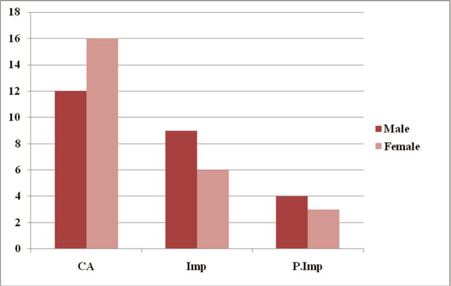
The results have been tabulated (Table 1 & 2) and the percentage difference in males and females in both the populations is shown in Graph 1 & 2.
| Subject | Male (n=25) | Percentage 100% | Female (n=25) | Percentage 100% |
|---|---|---|---|---|
| Congenital Absence | 12 | 48% | 16 | 64% |
| Impacted | 9 | 36% | 6 | 24% |
| Partly Impacted | 4 | 16% | 3 | 12% |
| Subject | Male (n=25) | Percentage 100% | Female (n=25) | Percentage 100% |
|---|---|---|---|---|
| Congenital Absence | 7 | 28% | 5 | 20% |
| Impacted | 8 | 32% | 7 | 28% |
| Partly Impacted | 10 | 40% | 13 | 52% |

- Prevalence of third molar eruption among South Indians. CA – Congenital Absence, Imp – Impacted, P.Imp – Partly Impacted.

- Prevalence of third molar eruption among Malaysians. CA – Congenital Absence, Imp – Impacted, P.Imp – Partly Impacted.
5. Discussion
Agenesis or a congenitally missing tooth is when a tooth fails to form between the ranges of age of its growth and development. This can also be called hypodontia. The third molar is a tooth that develops entirely after birth and is also the last tooth to erupt in all ethnic groups between the ages of 17–21 years despite racial variations in the eruption sequence. Environmental factors, systemic diseases, genetic polymorphisms, and teratogens were shown to affect tooth development with effects on tooth size, shape, position, and total absence. It is thus not surprising that aberrations in normal M3 patterning frequently occur, as this is the last tooth to develop. To date only a limited number of mutations of MSX1 and PAX9 have been proven to be associated with severe hypodontia in humans (Jacob, Prabhakaran & Mani, 2012). PAX9 is a transcription factor that is expressed in dental mesenchyme at initiation, bud, cap and bell stages of odontogenesis. Protein products of this gene serve as transcription factors that are responsible for the crosstalk between epithelial and mesenchymal tissues and are essential for the establishment of the odontogenic potential of the mesenchyme. The expression of PAX9 in the mesenchyme appears to be a marker for the sites of tooth formation. Mutations in this gene have been shown to be associated with autosomal dominant forms of oligodontia (agenesis of more than 6 teeth, MIM 604625) in humans (Jacob, Prabhakaran & Mani, 2012).
In a study done by Jacob et al. involving 734 Malaysians, 192 (26.2%) radiographs showed one or more missing third molars. Most patients with third molar agenesis had at least 2 missing third molars (19.3%). Agenesis of one or more third molars was more common in females (27.5%). The prevalence of M3 agenesis was highest among the Malaysian Chinese (32%) compared to the Malays (25.5%) and Indians (21.4%) (Jacob, Prabhakaran & Mani, 2012).
The impacted mandibular third molars are common amongst young adults. It was found that patients in the age group 21 and 25 years were most likely to present with impactions, with 68 (33.1%) patients, followed by patients between 26 and 30 years with 53 (26.2%). From this study, it is evident that impacted third molars decrease with corresponding increase in the age of patients. Furthermore, the study also showed that males between 21 and 25 years presented more frequently with impacted mandibular third molars than females. Obiechina et al. observed that patients in the 20–25 year age group presented with the highest number of impactions (Obiechina, Arotiba & Fasola, 2001).
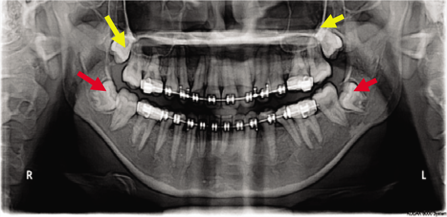
In South Indian Population, it was noted that only 48% of males and 64% of females have congenital missing third molar (Figure 1), and 52% of males and 36% of females have erupted third molar. 16% of males had partly impacted third molar (Figure 2) whereas in case of female it was 28%. Congenital missing third molars among females are 16% more than males (Graph 1). More males (36%) have impacted third molars than females (24%) (Table 1). It was also noted that majority of them have their maxillary third molars erupted before their mandibular third molars. In Malaysian Population, it was noted that only 28% of males and 20% of females have congenital missing third molars, and 72% of males and 80% of females have erupted third molars. 40% of males had partly impacted third molar whereas in case of female it was 52%. Congenital missing third molars among males are 8% more than females (Graph 2). More males (32%) have impacted third molars than females (28%) (Figure 3) (Table 2). It was also noted that majority of them have their mandibular third molars erupted before their maxillary third molars. In a study done by Jacob et al., stated that when compared to the maxilla, the mandible grows more than twice in length. However, unlike the Malay and Indian populations, Chinese patients exhibited very little third molar agenesis variation between the maxillary and mandibular arches. Among the Chinese, it was equally high in both arches (Jacob, Prabhakaran & Mani, 2012).

- Congenital Absence of Maxillary and Mandibular Third Molars.

- Partly Impacted Mandibular Third Molars.

- Impacted Mandibular Third Molars.
6. Conclusion
The prevalence of third molar agenesis in this study confirms its variations in relation to ethnic origin, gender, and location in the dental arch. It also confirms to the theory of the possible “extinction” of third molars in the future. This will have implications for future age-estimation studies and forensic identification. We recommend further studies on age-related dental and skeletal maturation among the various ethnic groups. It was also beyond the scope of this study to measure the skeletal and dental dimensions of the study population and determine their relationship with third molar agenesis, but such a study would have great relevance in dentistry, especially in understanding dental anomalies and management of orthodontic patients.
References
- (2007)Association between polymorphism in the promoter region (G/C-915) of PAX9 gene and third molar agenesis. Journal of Applied Oral Science: Revista FOB. ;15(5):382-386.
- [Google Scholar]
- (1987)Gradual change in human tooth size in the late pleistocene and post-pleistocene. Evolution. ;41(4):705-720.
- [Google Scholar]
- (2010)Evaluation of impacted mandibular third molars by panoramic radiography. International Scholarly Research Network. ;12(1):1-8.
- [Google Scholar]
- (2012)Third molar agenesis among children and youths from three major races of Malaysians. Journal of Dental Sciences. ;7(3):211-217.
- [Google Scholar]
- (1985). The effects of X-ray irradiation of the head region of eight-day-old rats on the development of molar and incisor teeth. Strahlentherapie. ;161(7):448-452.
- [Google Scholar]
- (2003)Genetic mapping of the absence of third molars in EL mice to chromosome. Journal of Dental Research. ;82(10):786-790.
- [Google Scholar]
- (2001)Third molar impaction: evaluation of the symptoms and pattern of impaction of Mandibular third molar teeth in Nigerians. Odonto-Stomatologie Tropicale. ;24(93):22-24.
- [Google Scholar]
- (2009). Impacted mandibular third molar surgery; an overview In: A publication by the Dentiscope Editorial Board. Vol 16. p. :22-24.
- [Google Scholar]
- (2009)Comparative study of third molar impaction in rural and urban areas of South–Western Nigeria. Odonto-Stomatologie Tropicale. ;23(90):25-28.
- [Google Scholar]
- (2005)Clinical evaluation of the effects of radiographic distortion on the position and classification of mandibular third molars. Dentomaxillofacial Radiology. ;34(2):96-101.
- [Google Scholar]
- (2003). The unresolved problem of the third molar: would people be better off without it? The Journal of the American Dental Association. ;134(4):450-455.
- [Google Scholar]
- (2004)Selectively preventing development of third molars in rats using electrosurgical energy. The Journal of the American Dental Association. ;135(10):1397-405.
- [Google Scholar]


