
Translate this page into:
Imaging Modalities for General Dental Practice - Bird’s Eye View
-
Received: ,
Accepted: ,
This article was originally published by Informatics Publishing and was migrated to Scientific Scholar after the change of Publisher.
Abstract
Imaging modalities employed in general dental practise include a plethora of intra and extra oral radiographic techniques for identification and diagnosis of various pathologies affecting the teeth and bones of maxillofacial region. The aim of this article is to highlight the application of the common and routine techniques currently available in the field of diagnostic radiology.
Keywords
Imaging
Intraoral Periapical Radiograph
Panoramic Radiograph
Radiograph

1. Introduction
With advanced diagnostic techniques, an undergraduate student would be clouded with the plethora of radio-graphic modalities currently available. While one can obtain theoretical knowledge of such newer techniques, the question arises, how does it apply in general practice? General dentistry is focused on diagnosis and management of patients which require treatment done within the scope of a dental clinic. The aim of this article is to describe the various radiographic imaging investigations a dentist can use for general dental practice (Figure 1). A brief outline of each radiographic investigations and the equipment required for set up with indications has been highlighted in the article.
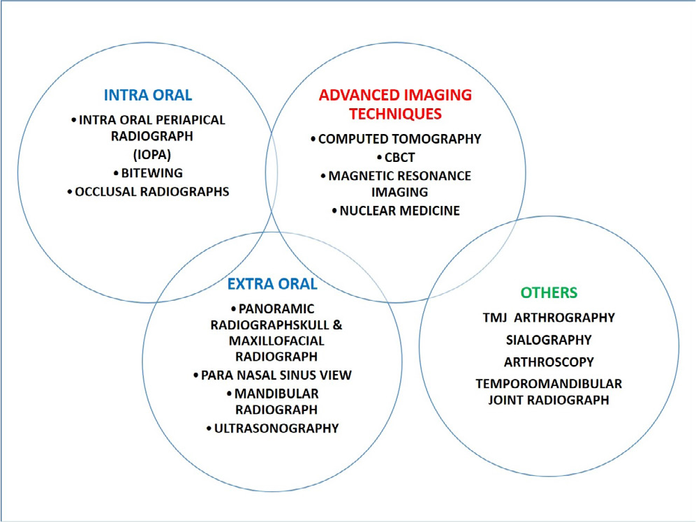
- Imaging modalities in dentistry.
2. Intra-Oral Radiographic Investigations
2.1 Intra Oral Periapical Radiograph (IOPA)
This is the most commonly used technique in general dental practice, convenient for patients and can be easily set up and maintained in a private dental clinic. Technically, this technique requires a simple protocol to be followed and can be easily mastered. The two common intraoral projection techniques used for radiographs are the paralleling technique and bisecting angle technique.
With this radiograph, a dentist can appreciate not only the teeth and also the surrounding hard tissues, alveolar bone and their apical portions. The exposure to the radiation is very minimal, easy to reproduce and provides exact representation of the tooth exposed. However, a few disadvantages are when the status of multiple teeth is required for examination, also prove difficult to apply in the patients with limited mouth opening and complying from children.
2.2 Bitewing Radiograph
This technique, also called interproximal radiographs, is easy to execute and cost effective for identifying interproximal caries. With this radiograph a dentist can visualise the crowns of the upper and lower arch and the alveolar crest. However, accurate reproduction can prove difficult and cone cut is frequently encountered. It does not require any specific angulations and discomfort compared to the patient as experienced by intraoral periapical radiograph.
2.3 Occlusal Radiograph
It is also known as Sandwich Radiography useful in locating supernumerary and impacted tooth within the maxilla or mandible. It is also used in assessing fractures of maxilla and mandible. Anatomical details cannot be appreciated and other defects such as overlapping and blurred images are encountered1. Two different angulations are required to enable dentist to view the maxilla and mandible (Table 1).
| MAXILLARY OCCLUSAL PROJECTIONS | MANDIBULAR OCCLUSAL PROJECTIONS |
|---|---|
| Upper standard or anterior occlusal | Lower 90˚ occlusal |
| Upper oblique occlusal | Lower 45˚ or anterior occlusal |
| Vertex occlusal, which is no longer used | Lower oblique occlusal |
3. Extra Oral Radiographs
In these radiographs, the films are placed outside the oral cavity, with the beam directed towards it. This type of radiography can be utilized for cases presenting clinically with large lesions, to study variations of the jaw, facial bones, to assess the growth of hard tissues, developmental defects, trauma and the temporomandibular Joint.
3.1 Panoramic Radiograph
This is the most commonly used extra oral radiographic modality in dental practice. It produces single tomo-graphic image of maxilla and mandible with shadows of supporting soft tissue structures. It provides a wide coverage of teeth and bones with comparatively minimal radiation exposure. One of the main advantages is its usefulness in patients with trismus. Patient cooperation is minimal and the radiographs are easy to interpret. This radiography requires an initial investment and images do not provide sufficient detail for periodontal or endodontic procedures often with overlapping of anatomic structures.
3.2 Cephalometric Radiograph
This radiography is mostly used in Orthodontics to study the relationship of teeth to the jaw and jaws to the rest of skeleton for treatment planning and orthognathic surgery. For this radiography, the equipment utilized is available with panoramic radiography and differs in head positioning.
3.3 Skull and Maxillofacial Radiograph
This type of radiography, is used to diagnose trauma or lesions of the skull or maxillofacial region which cannot be detected using a panoramic radiography. The technique, principles, interpretation and setup of this radiography exceeds the regular usage in a private dental practice and is the choice of the dentist to invest time and money on this particular radiography.
If at all cases of suspected fracture of maxillofacial region do report, they can be referred to hospital with mention of the type of radiograph to be taken. The types of radiograph and its indications are given in Table 2.
| S.No | TYPES | FILM SIZE | INDICATIONS |
|---|---|---|---|
| IOPA | 0 Size – 22 x 30 mm (For children) | Identification of apical infection or inflammation To determine the periodontal status. Trauma to the teeth and alveolar bone. | |
| 1 Size – 24 x 40 mm (For adults anterior) 2 Size - 31x 41mm (For adults posterior) |
To assess the tooth eruption, planning extractions, during endodontic treatment. For planning implants. |
||
| Bitewing | Size 0, (Same size as IOPA) Size 2 | To identify caries in proximal sides. To record restorations To keep a check on progress of dental caries To examine the periodontal status | |
| Occlusal | Size – 57 x 76 mm | To locate supernumerary impacted tooth To identify dentoalveolar fractures. |
3.4 Radiography of Mandible (PA Mandible)
Among the extra oral radiographs, this type of radiography is commonly used to diagnose fractures of posterior third, angle of the mandible as well as mandibular deformities. The three types are
PA Mandible - Posterior anterior view of mandible and ramus.
Rotated PA Mandible - One side of the mandible.
Lateral oblique - Anterior body of the mandible, Posterior body of the mandible, Ramus of the mandible in oblique angulations.
3.5 Radiography of Temporomandibular Joint
The most commonly used technique like panoramic radiography and other extra-oral radiograph cannot help in arriving to a specific diagnosis emphasises the relevance of temporomandibular radiography. This technique helps to examine the TMJ bony relations and give a complete detail about the articular surface, condylar head and neck and the walls of temporomandibular joint and their supporting structures and bone also their defects. The types of TMJ radiography commonly used are Transcranial, Transpharyngeal, Transorbital and Reverse Town Projection2.
A summary of type of extra oral radiography and its indications is given in Table 3.
| S.No | TYPES | SIZE OF FILM | INDICATION |
|---|---|---|---|
| 1. | PANORAMIC RADIOGRAPHY (OPG) |
5 x17 or 6 x12 inch |
Require extensive coverage of jaws. •To assess fractures, due to trauma and impacted teeth and unerupted teeth. •For Odontogenic lesions, such as cyst and tumours. |
| 2. | Other than OPG | 8 x 10 or 5 x 7 inch |
• To study of large lesion, jaw, facial bones and their developmental defects. • Examination of skull vault and jaws • Examination of sinuses • Examination of bony defects in skull and maxillofacial region • For the different types of treatment planning and evaluation of skull & maxillofacial region. |
4. Advanced Imaging Technique
4.1 Computed Tomography (CT)
Unlike other methods, Computed Tomography gives a clear picture of extensions of intracranial defects such as tumours, haemorrhage and trauma of the maxillofacial region. It also helps to assess boundaries of cysts and bone lesions, osteomyelitis and even pathologies of the TMJ. Although useful in preoperative study of implants, a preferred alternative would be Cone Beam Computed Tomography (CBCT). It produces a sectional or slice images using crystal or gas electrons converting the beam into a digital data. The image produced by CT provides fine differentiation of soft and hard tissue enabling dentists to appreciate features that would be missed in extra-oral radiography and can be manipulated such as reconstruction and enhanced, but the equipment used is expensive, technique sensitive and requires patient cooperation. A point worth noting that metal objects (e.g., Bone plating in fractures) can destruct the image3.
4.2 Cone Beam Computed Tomography (CBCT)
This is a modified and refined technique of CT providing 3D imaging of maxillofacial structures. Compared to CT, the radiation exposure is low and provides accurate and intricate details of the hard tissue. Based on the sensor and software, and type of practice, dentists can invest on their requirements thus reducing the overall cost of the equipment4.
4.3 Magnetic Resonance Imaging (MRI)
In addition to other methods, this imaging modality enables specific diagnosis about soft tissue conditions, intra cranial lesions, soft and hard tissues, tumours jaw and TMJ. It works in principle of magnetic field and radio-frequency. When the patient is placed in magnetic field the atoms of the body emits radiofrequency pulse which leads to absorption of energy and is used to detect and record the images in the scanner. Therefore, no exposure to radiation is required and the image produced is of high resolution. However, it is contraindicated in patients with surgical clips, cardiac pacemakers, cochlear implants, first trimester of pregnancy5.
5. Other Imaging Techniques and Nuclear Medicine
Other advanced techniques makes use of collimated beam radiation produce images of greater quality and details, they are very much useful in scanning posterior-anterior maxillofacial complex. Advanced methods such as, Nuclear medicine makes use of radionuclide label traces, detect early disease and also specified growth of lesion and extent of its borders. Some technique permits each processed at a rapid rate to a long motion. Unlike other radiographic technique, the above mentioned technique helps in identifying minute defects of soft and hard tissues of the skull and maxillofacial region.
Ultrasound is one modality that is utilized to examine pathologies related to salivary glands. These are not routinely used in a private practice and require training for its usage6.
6. Conclusion
This article outlines the basic set up of the routinely employed radiographic modalities with focus on its advantages and disadvantages, and indications for use. The choice of instrument for a private dental clinic depends on the nature of practice and the monetary investment. Advanced imaging modalities serve as additional information which may or may not be necessary in treatment but its use is justified if it outweighs the higher radiation exposure for enhanced quality of treatment.
References
- MRI in dentistry-A future towards radiation free imaging-systemic review. Journal of Clinical and Diagnostic Research. 2016;10(10):1-2.
- [Google Scholar]
- Nuclear medicine in orofacial diagnosis: A review. Journal of Medicine Radiology and Surgery. 2016;2(4):1-3.
- [Google Scholar]


