Translate this page into:
Aquaporins in Salivary Gland - The Water Fa(u)cet of an Acini?
*Author for correspondence
-
Received: ,
Accepted: ,
This article was originally published by Informatics Publishing and was migrated to Scientific Scholar after the change of Publisher.
Abstract
Salivary glands are exocrine glands secreting saliva into the oral cavity. The primary function of the saliva is to protect and hydrate the mucosal structures of the oral cavity. The lubrication and hydration of the oral mucosa is provided by the water content of the saliva which forms approximately 99% of its composition. Aquaporins are water channels expressed in acini of salivary glands and play an important role in formation of saliva. Aquaporins are transmembrane water permeable proteins involved in transcellular water flow. In addition to being permeable to water, some Aquaporins can be permeable to small solutes, including cations, glycerol and gases. The present article reviews the basic histology of salivary gland, its ductal system and also physiology of secretion of saliva and highlights the role of Aquaporins in saliva formation.
Keywords
Aquaporins
Clinical Application
Expression
Localization
Salivary Gland
Structure
1. Introduction
Saliva is a mixture of fluid secreted by salivary gland. Based on the size, there are two groups of salivary glands present in the human body, i.e., major and minor salivary glands. The major salivary glands are the Parotid gland, Submandibular gland and Sublingual gland. These are located outside from the oral mucosa the secretions of which are emptied into the oral cavity by the ductal openings. The minor salivary glands are those that are found in the oral cavity, examples are the Von Ebner gland, labial gland, palatine gland etc. Based on the viscosity and secretory acini there are three types of secretions, i.e., serous secretion, mucous secretion and mixed secretion. The role of Aquaporin is to permit the water inflow which determines the viscosity and tonicity of the saliva secreted.
2. Ductal System of Salivary Gland
Salivary glands are composed of a parenchymal component, each consisting of the secretory units and the ductal system. The secretory units consist of the acinar cells, which are the basic structural and functional unit of a gland. They are responsible for the synthesis and secretion of saliva.
Microscopically, the ductal system consists of three parts, i.e., the intercalated duct, striated duct and the excretory duct. The intercalated duct and striated duct, along with the secretory unit are the main components of an intra-lobular duct. Another inter-lobular duct consists of the excretory ducts of a gland1,2. All these ducts function to transport saliva secretion through the terminal excretory duct and into the oral cavity (Figure 1).

- Ductal System of Salivary Gland.
3. Physiological Mechanism of Saliva Secretion
Saliva secretion mainly occurs in two stages, the first is to produce isotonic plasma-like primary saliva and secondly modification of the primary saliva in ductal system.
Isotonic plasma like fluid (rich in NaCl) is initially secreted by the acinar cells. Where it involves few ionic transporters for the transportation of the Na+ and Cl- across the basolateral membrane. Once the Na+ and Cl- are secreted into the lumen more than its equilibrium (hypo-tonic) in comparison to intracellular concentration, water gradient is produced. This causes influx of water into the lumen from the intracellular with the help of aquaporins (AQP5) present in the apical membrane of acinar cells. Primary saliva is produced.
As mentioned above the ductal system is composed of three types of ducts; intercalated, striated and excretory ducts. Each duct plays an important role in modification of composition of the primary saliva. This modification starts with the reabsorption of the Na+ and Cl- by ductal cells with the presence of K+ and HCO3- as the exchanger of the ions respectively1–3. This reabsorption and secretion of the ions require ion channels and transporters (Figure 2).
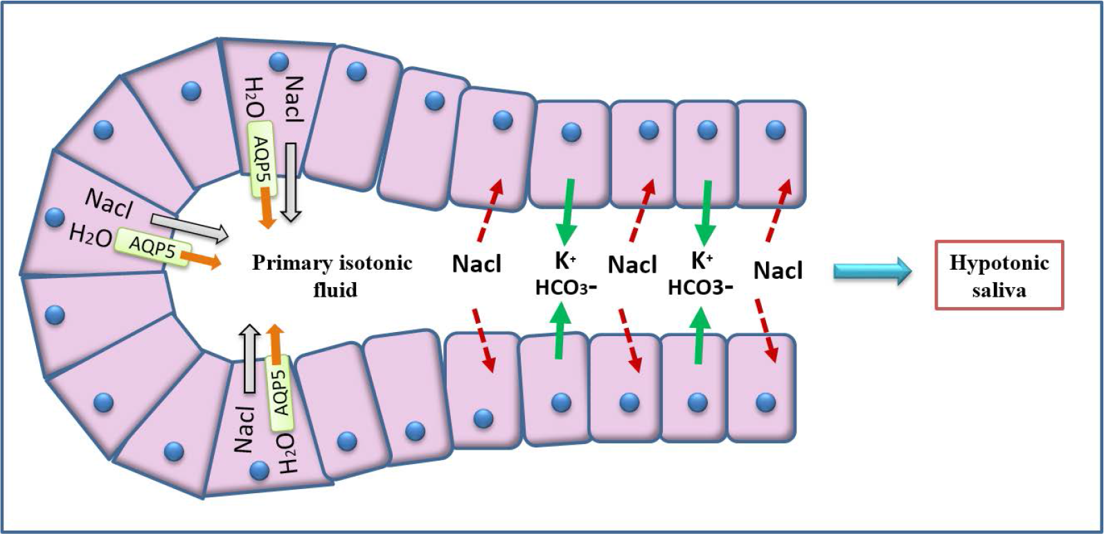
- Physiology of Saliva Secretion.
4. Aquaporins
4.1 History
Before year 1992, scientists believe that water in our body only transported by osmosis and diffusion. In 1992, scientist Peter Agre discovered aquaporin and he won a noble prize in 2003.
4.2 Structure, localization and Expression of AQP
Aquaporins are transmembrane structures which exists as monomer consists of six helices and five loops. With it being a type of protein, it has N and C terminals which are located intracellularly.
There are 13 types of aquaporins found in our whole body for regulation of water homeostasis. In salivary glands, only three types are present which are AQP1, AQP3 and AQP5. AQP1 is present in the myoepithelial cell, AQP3 on the basolateral membrane and AQP5 on apical membrane4 (Figure 3, 4, 5).
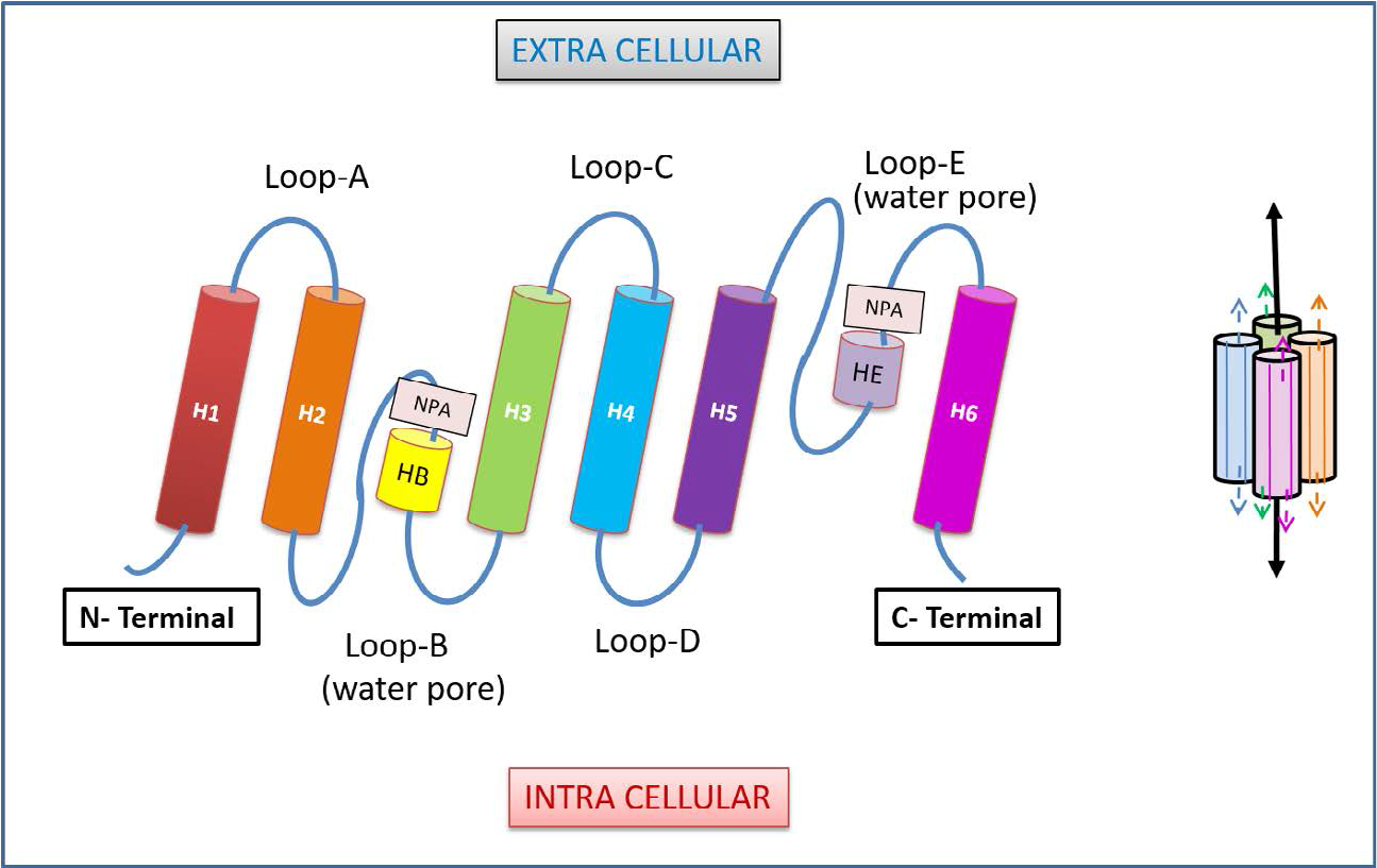
- Structure of Aquaporins.
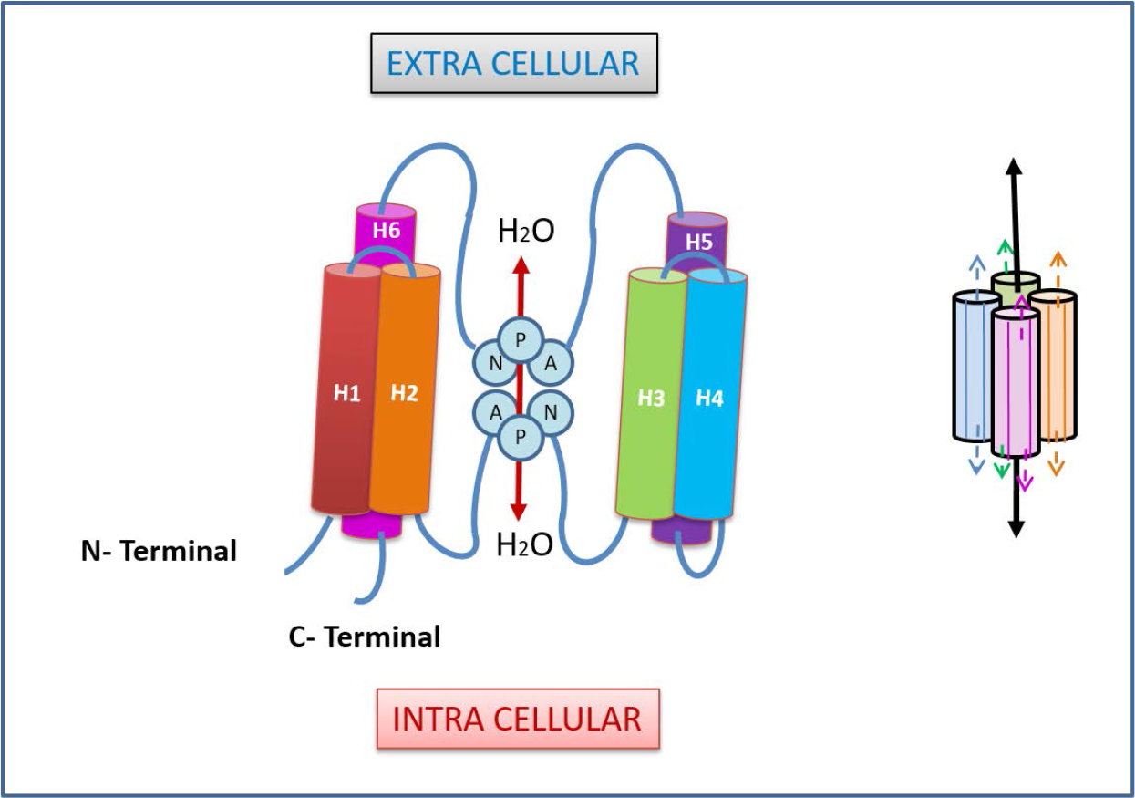
- Arrangement of Aquaporin Molecule.
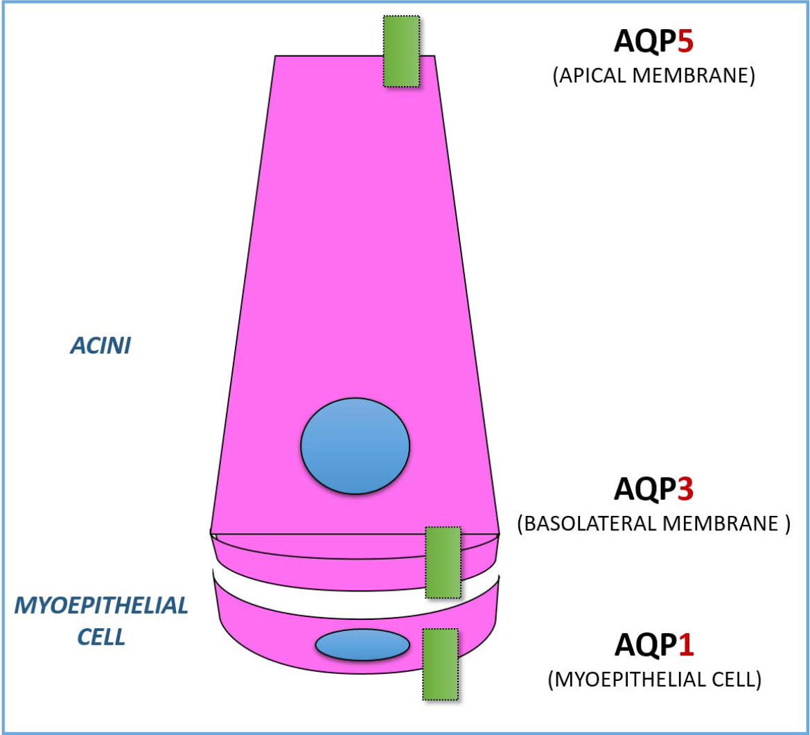
- Expression and Localization of Aquaporin in Acini.
4.3 Role of Aquaporin in Salivary Gland
Since 1992, few experiments have been conducted to find out the role of aquaporin. The most significant experiment is that experiment targeting knockout mice in which their AQP5 in salivary glands have been removed.
A drug named pilocarpine had been given to the mice to regulate the transmembrane AQP5. The resulting saliva secretion of the mice has reduced by 60% when compared to the normal secretion. On the other hand, water permeability of saliva also decreased which is 65% in parotid gland and 77% in sublingual gland. These experimental findings showed that saliva secretion is more viscous and hypertonic when compared to normal saliva which is hypotonic suggesting the regulatory function of aquaporins in water permeability in the formation of saliva5,6.
4.4 Role of AQP5 Trafficking in Saliva Secretion
The water molecules move from extracellular fluid into the lumen from basolateral membrane to apical membrane of acini cell. The mechanism of intracellular trafficking of AQP5 shows how the water is transported.
This trafficking is initiated by acetylcholine and nor-adrenaline binding to their receptors which are the M3 receptor and β receptor resulting in the increase in intra-cellular calcium and cAMP. Following which Calcium and cAMP will then activate protein kinase C, calcium-calmodulin-dependent protein kinase and protein kinase A in order to induce the phosphorylation of AQP5 present in the intracellular vesicles. It will further lead to their fusion with apical membrane. Once the phosphorylated AQP5 fuses with apical membrane, water molecules are allowed to flow into gland lumen6,7 (Figure 6).
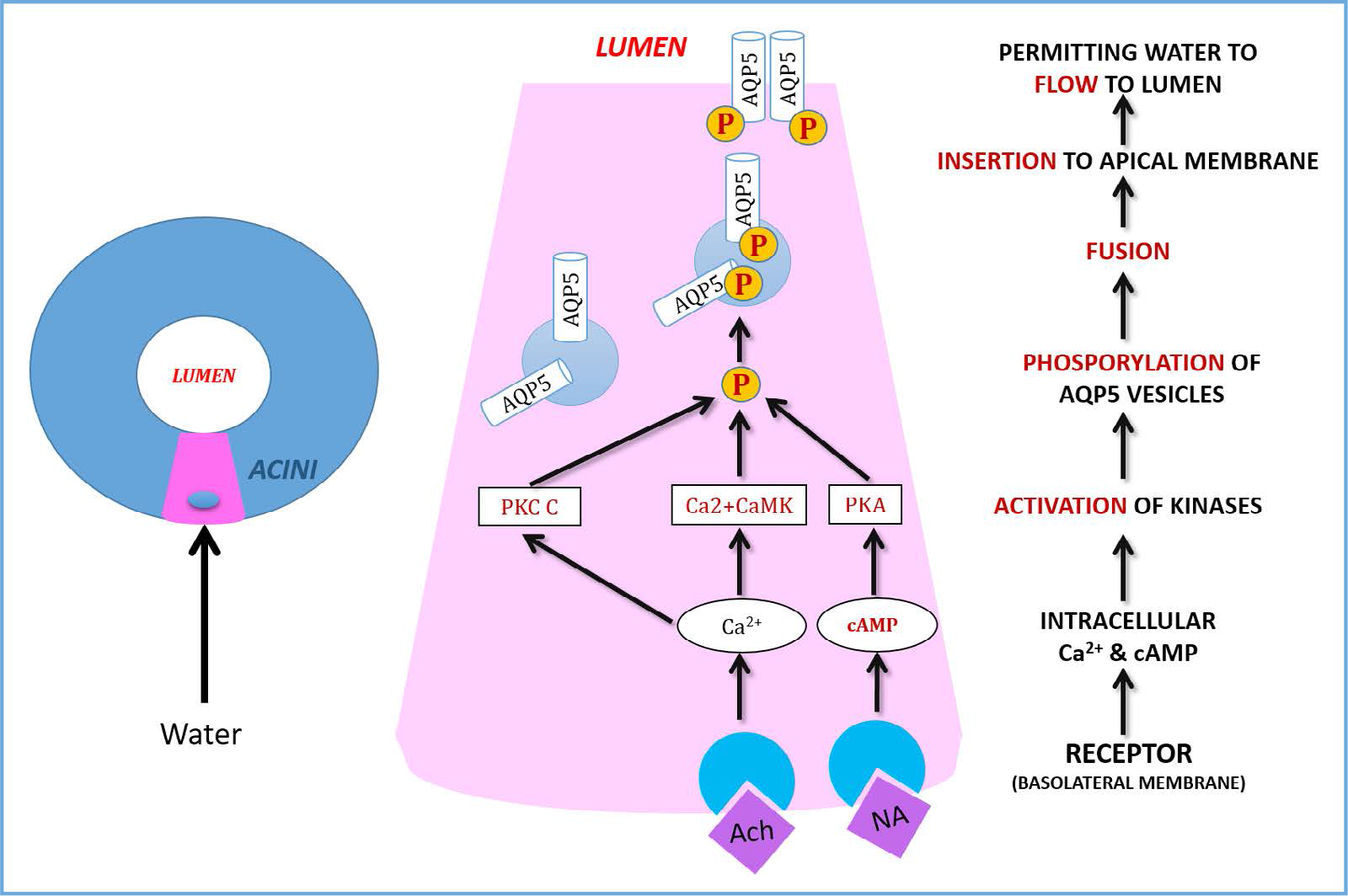
- Role of AQP5 Trafficking in Saliva Secretion.
5. Clinical Application
In normal secretion, humans secrete 750 to 1000mL of saliva per day. If the individual have reduction of secretion to about 50%, it is considered as Xerostomia. The role of Aquaporins in few clinical conditions have been discussed below8.
5.1 Sjögren’s Syndrome
Sjögren’s syndrome is an autoimmune disease which affects predominantly women. Pathologically, it is characterized by the dysfunction of salivary glands resulting in the loss of saliva secretion. The main cause of this disease is the abnormal localization of AQP5 at the basolateral membrane of acinar cells instead of at the apical membrane. Besides, it is also believed that it is due to an altered gene expression of AQP5. There are two types of Sjögren’s syndrome, which are the primary type and secondary type. The primary type occurs alone whereas the secondary type is associated with other autoimmune diseases e.g., rheumatoid arthritis.
5.2 Radiation Therapy
Radiation therapy is one of the treatment that is received by cancer patients with the help of chemotherapy. However, this radiation therapy leads to xerostomia due the damage of the acinar cells in the ductal system. Aquaporins will be lost or decreased in the ductal system of salivary gland which results in altered saliva tonicity.
5.3 Senescence
Saliva secretion will gradually decrease as human senescence, which is the state of being old or in the process of aging. Thus, it is said that senescence is also one of the reasons of xerostomia.
References
- Chloride channels and salivary gland function. Crit Rev Oral Biol Med. 1999;10(2):199-209.
- [CrossRef] [PubMed] [Google Scholar]
- Distribution and roles of Aquaporins in salivary glands: A review. Biochimicaet Biophysica Acta. 2006;1758:1061-70.
- [CrossRef] [PubMed] [Google Scholar]
- Identification and localization of aquaporins water channels in human salivary glands. Am J Physiol Gastrointest Liver Physiol. 2001;281:G247-G257.
- [CrossRef] [PubMed] [Google Scholar]
- The salivary gland fluid secretion mechanism: Mini-review. The Journal of Medical Investigation. 2009;56:192-6.
- [CrossRef] [PubMed] [Google Scholar]
- Role of aquaporins in saliva secretion: A review. OA Biochemistry. 2013;1(2):1-6.
- [CrossRef] [Google Scholar]
- Aquaporins in salivary gland: From basic research to clinical applications: A review. International Journal of Molecular Science. 2016;17(2):1-13.
- [CrossRef] [PubMed] [Google Scholar]


