Translate this page into:
An Interesting Case of Lower Lip Cleft with Syndromic Features
-
Received: ,
Accepted: ,
This article was originally published by Informatics Publishing and was migrated to Scientific Scholar after the change of Publisher.
Abstract
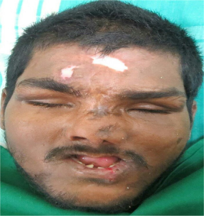
Observation of a patient named Sanjay, a 23 year old male, revealed a case of anatomical curiosity, an uncommon variant of a developmental anomaly and masochistic habit, an atypical median mandibular cleft with distinctive facial appearance and intellectual disability. Facial gestalt is characterized by a round face, the heavy horizontal eyebrows with narrow palpebral fissures.
Keywords
Mental Retardation
Over Growth Syndrome
The Median Mandibular Cleft
Tessier 30 Cleft
1. Introduction
According to Dr. Paul Tessier (1976), Clefts may include mouth, eyes, cheeks, ears and forehead which continues to the hairline. These cranio facial clefts are referred to as “Tessier cleft’s”. They are numbered from 0-14 to indicate the location and the extent of the cleft using the mouth, nose, eye sockets as landmarks with the midline designated as “0”. The extensive conditions are “OROOCCULAR CLEFT” and “FRONTONASAL DYSPLASIA”1 (Figure 1).

- Tessier classification of orofacial cleft 1976.
“Tessier 30 cleft, or Lower midline facial cleft”, also known as “Median mandibular cleft” is a uncommon anomaly. The median cleft of the mandible was reported by Couronne in 1819 and Dr. Paul Tessier in 1976”2. Erdogane et al., (1989) did another literature review and found 48 patient with the midline cleft of the lower lip of these, 37 involved mandible3.
2. Case Report
A 23 years old male reported to the department of oral maxillofacial surgery Vinayaka Mission Sankarachariyar Dental College, Salem, Tamil Nadu, with a chief complaint of drooling of saliva and deformity of his lower lip. He had no family history of a similar disorder. His parents had consanguinous marriage and his mother did not give a history of taking any medications or exposure to radiation during her pregnancy. Physical examination showed a visible midline cleft of the lower lip with the drooling of saliva (Figure 2). The lower alveolus and the tongue are normal on inspection and palpation, a notch was felt on the mandible (Figure 3). The patient had masochistic habit and blindness and loss of digits on the hand and feet , the scars on the forehead and cleft of the midline mandible, deviated nasal bridge and absence of maxilla and premaxilla.
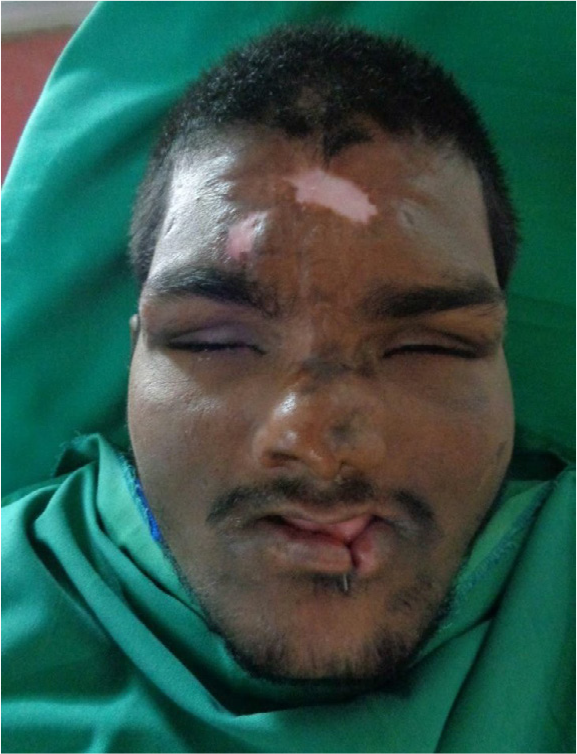
- Preoperative extraoral picture shows lower lip.
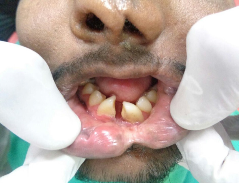
- Preoperative intraoral picture.
3. Discussion
The midline cleft of the lower lip is a midline vertical cleft of the soft tissue of the lower lip. It is more commonly accompanied by a cleft of the mandible. This disorder is not hereditary and has no sex predilection. The frequency is 4 to 5 cases per million births. Gorlin et al., (1990) said that in nearly all cases there is ankyloglossia, where the tongue tip is attached to the median cleft by a frenum. The cleft may be severe, bifurcating the mandible, the tongue, and the structures of the mid neck down to the hyoid bone4. Ranta (1984) described 12 case of the minimal cleft of the lower lip of which was a cleft palate in 7 cases, Robin sequence in 3, and agenesis of the incisors in 2 patients5. Embryologically, mandible develops from the cartilage of first pharyngeal arch, which is present at 5th week. The cartilage of the first arch consist of a small dorsal portion, the maxillary process, which extend forwards beneath the region of the eye, and a much larger ventral portion , the mandibular process or Meckel’s cartilage. The mandible first appears as a mesenchymal condensation external to the cartilage. Ossification commences in this condensation between 5th and 6th week in the region near the mental foramen, retrogresses and becomes surrounded and invaded by the membranous ossification of the mandible. As the ossification spreads, the inferior alveolar nerve is surrounded and the two centres of ossification migrate towards each other. Endochondral ossification occurs in ventral tip of the meckle’s cartilage forming the mental ossicles that are later incorporated into mandible.
Median mandibular cleft arise from a failure of the mesodermal penetration. This results of under development of the ventral tip of meckel’s cartilage that leads to a failure of ossification and the production of the cleft.
A midline defect bony measuring 8*15mm (width*depth) is seen in the midline in the mental region of mandible (region of incisors) with associated focal defect in upper and lower lip left parasagittal location.
The lower left and right central and lateral incisors are not visualized .lower canine, premolar teeth. The lower first, second and third molar teeth appear normal. No evidence of any radiolucency around the root. Rest of mandible is normal (condyl, ramus and body).
Bilateral maxillary sinuses are rudimentary with associated mucosal thickening. All other sinuses are clear. Mild deviated nasal septum to left side is seen. Nasal bone is normal. Bilateral temporo mandibular joint appears normal. No fracture seen in facial bones. Visualized orbits show atrophied and deformed bilateral globes with hyper dense foci in it suggestive phthisis bulbi. Orbital bones are normal. Visualized brain parenchyma are normal.
4. Surgical Procedure
Cleft of the lower lip was repaired under local anesthesia. A triangular vermilion flap was reflected. Orbicularis oris muscle was dissected by using iris scissor. Orbicularis oris muscle was reoriented and sutured with 4.0 vicryl.
Triangular vermilion flap was transposed and sutured by using 5.0 prolene (Figure 6, 7 and 8). The skin was sutured with 5.0 prolene (Figure 9). Postoperatively the wound healing was satisfactory (Figure 10). Through this surgical correction of the cleft, the patient’s functional problem of salivary drooling had been corrected.
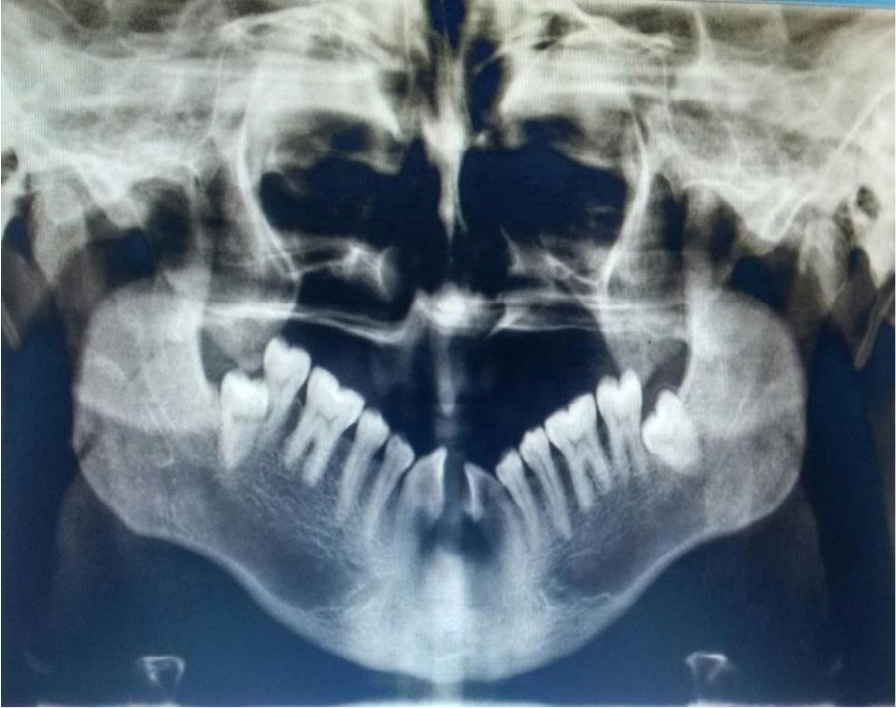
- Orthopantamogram shows complete absence of the maxillary teeth, and the mandibular central incisors and lateral incisors. The cleft involving midline of the mandible and small notching is present on mandibular alveolous with mesial drift of the remaining teeth.
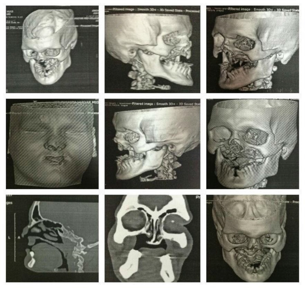
- Computer tomography of facial bone shows non visualization of maxilla and its alveolar process and all upper teeth .Palatine process of maxilla is poorly visualized with defect in visualized posterior part of hard palate and communication between oral and nasalcavity.
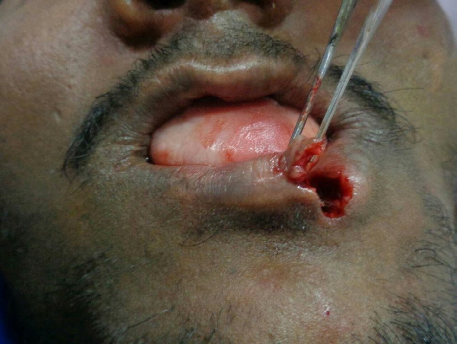
- Under local anesthesia 1:80000 infiltrated in the lower lip by using No:11 blade vermilion border is desected, triangular shape vermilion flap is reflected.
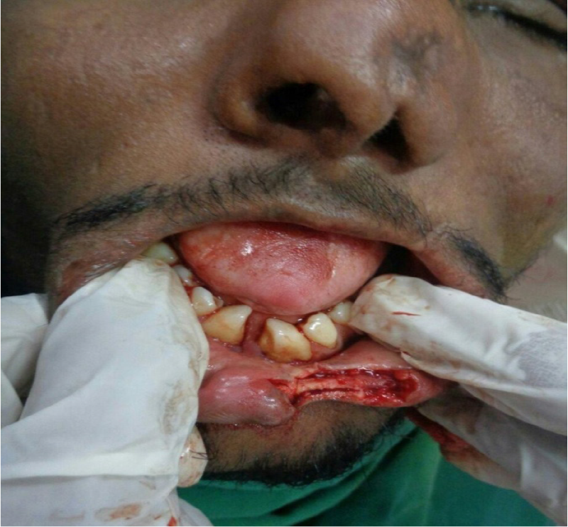
- Orbicularis oris muscle is dissected by using iris scissors.
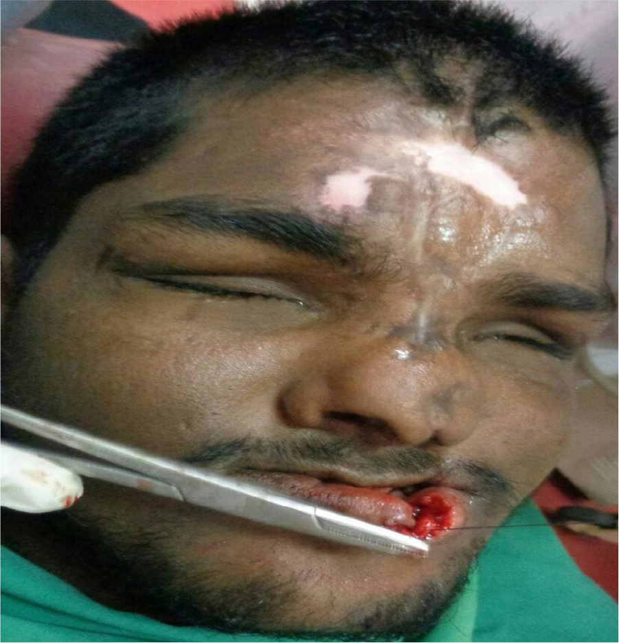
- Orbicularis muscle is reoriented and sutured with 4.0 vicryl.
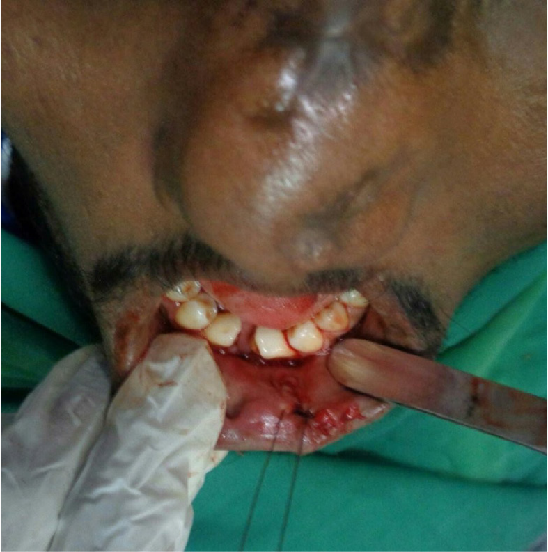
- Triangular vermilion border sutured on opposite side by using 5.0 prolene.(all corners of the flap sutured first) skin is suture with 5.0 prolene.
- Orbicularis muscle is reoriented and sutured with 4.0 vicryl.
References
- Anatomical classification of facial. Cranio-Facial and Latero-Facial Clefts. Journal of Max illofacial Surgery. 1976;4:69-92.
- [Google Scholar]
- Rare craniofacial clefts In: Mc Carthy JG, ed. Plastic Surgery. W.B. Saunders; 1990. p. :2940.
- [Google Scholar]
- Incomplete median cleft of the lower lip with cleft palate, the Pierre Robin Anomaly or hypodontia. Int J Oral Surg. 1984;13:555-8.
- [Google Scholar]


